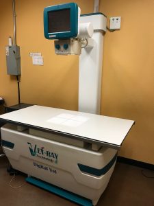
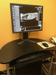
Many people are familiar with radiographs (x-rays) through personal experience or through their pet having them done. It is a very common procedure done in pets to diagnose problems such as broken bones, masses (tumors), obstructions, bladder stones, or hip dysplasia. X-rays were discovered in 1895. Since then, they have become a valuable tool in the diagnosis and treatment of disease.
Radiographs rely on the different densities of various parts of the body. The tissue images that show up on the radiograph consist of white, black, and shades of gray. The more dense the body part such as bone, the whiter it appears on the x-ray. The less dense the body part such as the lung filled with air, the blacker it appears on the x-ray. Muscle and internal organs show up as various shades of gray depending on the density. A contrast media (dye) can be injected into the patient as with a myelogram, or given to the patient as with a barium series to provide further information. With experience reading it, the veterinarian is able to use the radiograph to help make a diagnosis.
When using radiographs to assist in diagnosing a condition, two views should always be taken. This is because the superimposition of one body part over another can hide a problem or cause a misinterpretation of the radiograph. The second view is usually at a 90º angle from the first. As an example, looking at only the first (top) view of the chest of a cat, as shown on the right, it may appear as though the cat has been shot in the heart. When looking at the second (bottom) view, we now see the lead is just under the skin.
Depending on the body part being x-rayed, the patient may be able to remain awake. Sedation or anesthesia may be needed if the animal is painful, aggressive, or will not lie still, since movement of the pet will cause blurring of the radiograph, making it, in many cases, worthless. Artifacts can occur if any object is between the x-ray machine and the x-ray plate. Collars and tags need to be removed before x-rays of the head/neck/shoulder region are done. Some soft tissue such as the brain, spinal cord, and tendons can not be viewed on x-rays and other types of imaging are necessary.
http://www.peteducation.com/article.cfm?c=2+2116&aid=1013


